




Frequent glass prescription changes
Keratoconus: Protect your cornea, preserve your vision
Keratoconus can slowly change the shape of your cornea and cause distorted or blurred vision. Early diagnosis and timely treatment can help control progression and improve visual quality.

What is Keratoconus?
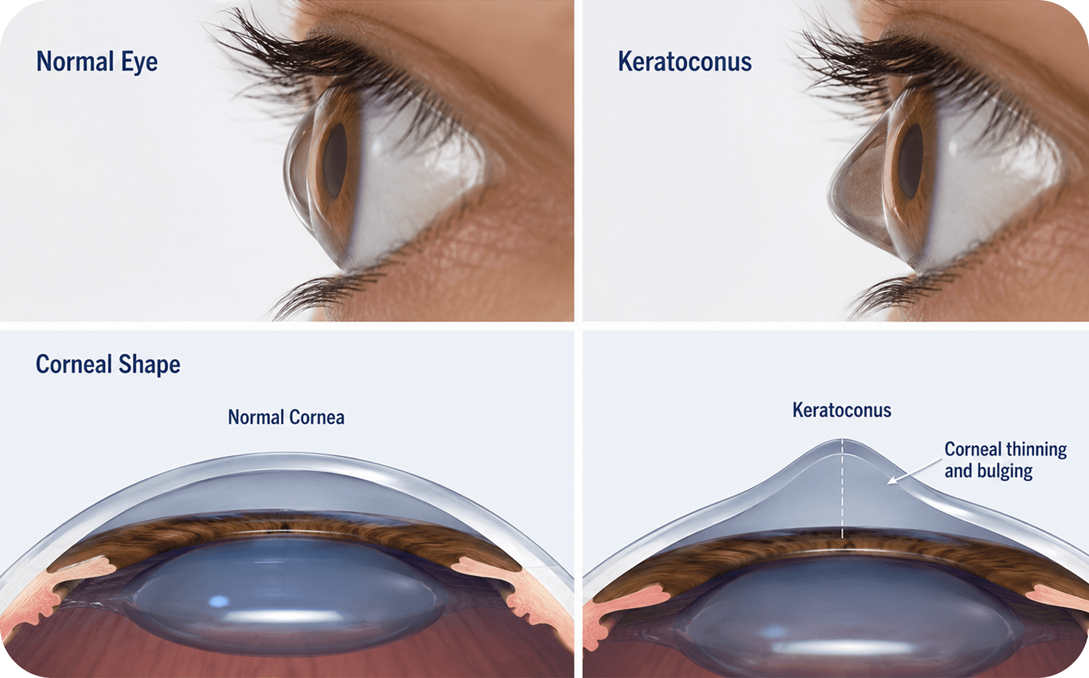
Keratoconus is a condition where the normally round cornea becomes thin and gradually bulges into a cone-like shape.
This irregular shape affects how light enters the eye, leading to blurred, distorted, or fluctuating vision.
It often starts with frequent changes in glass power, increasing astigmatism, glare, halos, or difficulty seeing clearly even with spectacles.
A detailed corneal evaluation is needed to diagnose the condition and understand whether it is stable or progressing.
Symptoms you may notice
Keratoconus can be missed in the early stage because it may look like a simple change in glass power.
What can worsen Keratoconus?
Some habits and conditions can increase the risk of corneal weakening or faster progression.






Corneal Thickness Check
Measures how thin the cornea is and helps decide treatment safety.

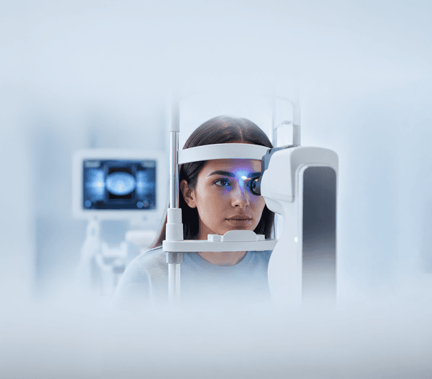
Corneal Topography / Tomography
Creates a detailed map of the cornea and helps detect early irregularity.
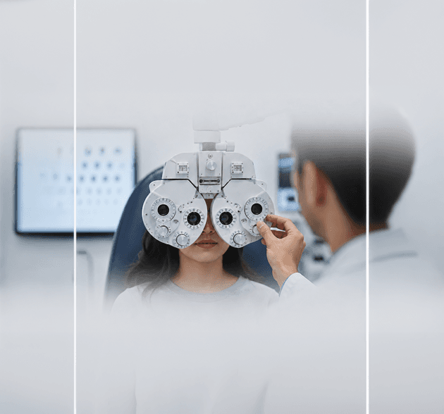
Refraction Test
Checks spectacle power and irregular astigmatism.
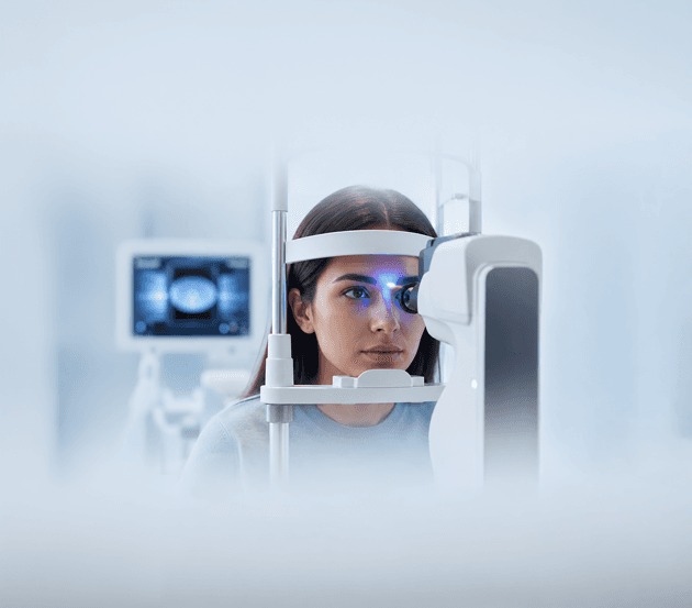
Corneal Thickness Check
Measures how thin the cornea is and helps decide treatment safety.
Corneal Topography / Tomography
Creates a detailed map of the cornea and helps detect early irregularity.
Our Keratoconus treatment approach
At Pixel Eye Hospitals, keratoconus care starts with a detailed corneal evaluation.
The goal is to understand whether the condition is mild, moderate, advanced, or actively progressing.
Treatment is planned with two priorities: first, to slow or stop further progression of the disease; second, to improve vision quality using the right glasses, contact lenses, or surgical options where required.
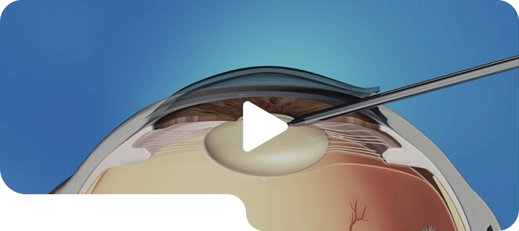
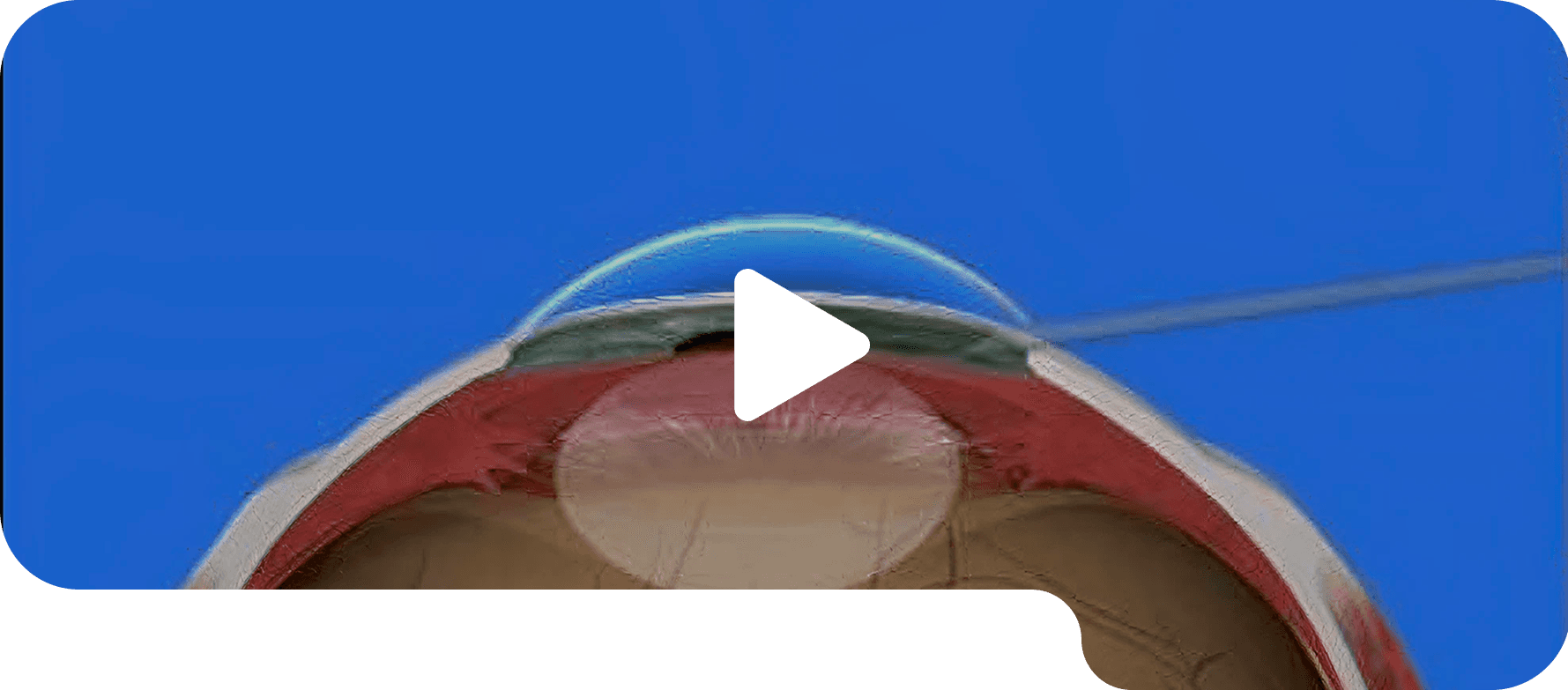

1. Glasses & Soft Contact Lenses
Glasses or soft contact lenses may help in early or mild keratoconus when vision can still be corrected comfortably.

2. RGP, Hybrid & Scleral Lenses
Special contact lenses may be recommended when regular glasses do not provide clear vision because of corneal irregularity.
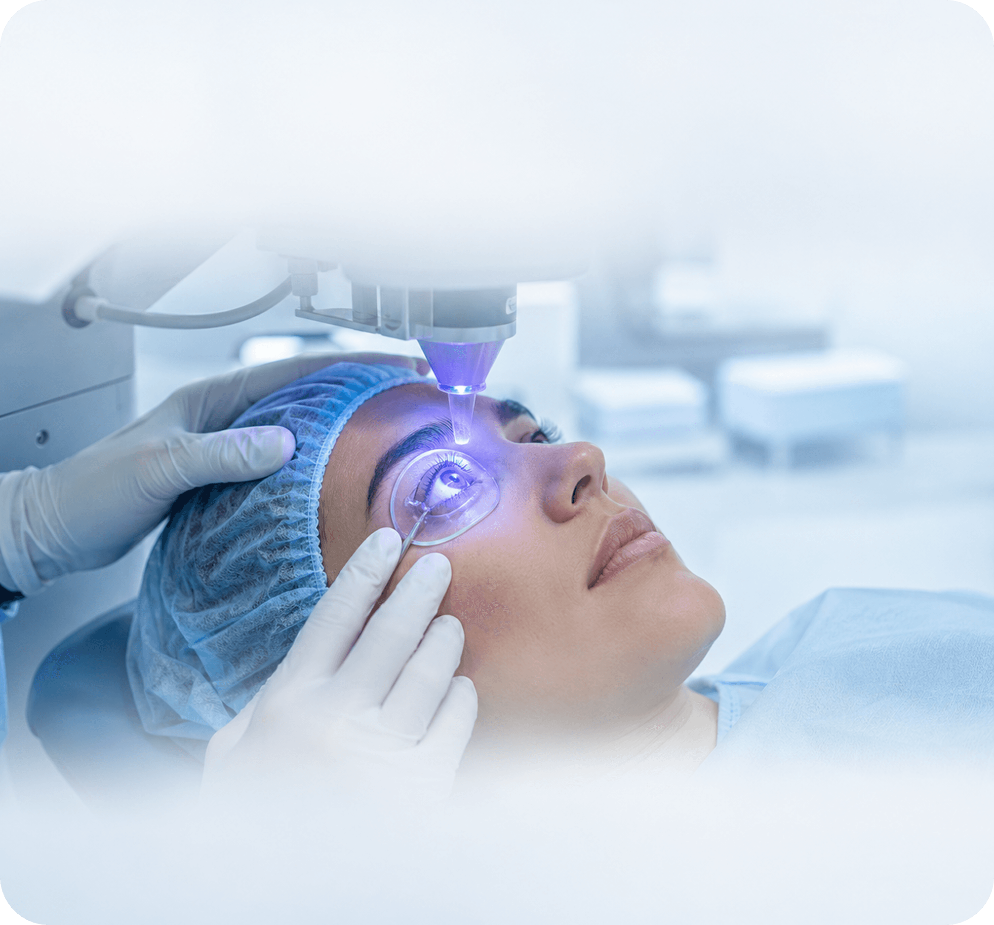
3. Corneal Collagen Cross-Linking / C3R
C3R helps strengthen the cornea and is commonly advised when keratoconus shows signs of progression.

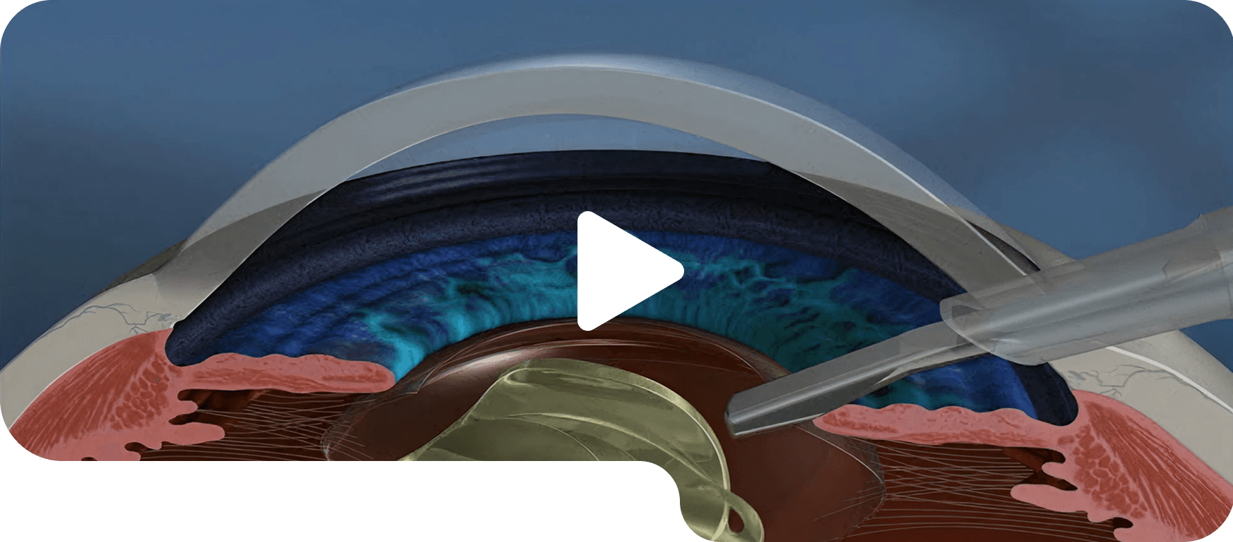
4. Intracorneal Ring Segments
Small ring segments may be placed inside the cornea in selected mild to moderate cases to help flatten the cornea and improve vision.
1. Glasses & Soft Contact Lenses
Glasses or soft contact lenses may help in early or mild keratoconus when vision can still be corrected comfortably.
2. RGP, Hybrid & Scleral Lenses
Special contact lenses may be recommended when regular glasses do not provide clear vision because of corneal irregularity.
Clinical expertise
Keratoconus care at Pixel Eye Hospital is led by experienced ophthalmologists trained in advanced



Dr. Krishna Poojita
MBBS, DNB (Ophthalmology), MRCS (Edinburgh), FICO (UK), FPRS (Narayana Nethralaya) Cataract & Refractive Surgeon
View Doctor Profiles
FAQs

For detailed and personalised guidance on keratoconus diagnosis, corneal cross-linking, specialty contact lenses, or advanced treatment options, schedule a consultation with our cornea specialists. Early diagnosis and timely treatment can help preserve your vision and reduce the progression of keratoconus.
Keratoconus is an eye condition in which the normally round cornea gradually becomes thinner and bulges into a cone-like shape. This irregular shape causes blurred and distorted vision that usually worsens over time if left untreated.
Early symptoms may include blurred vision, frequent changes in spectacle power, increased sensitivity to light, glare while driving at night, ghost images, and difficulty seeing clearly even with glasses.
Keratoconus cannot be completely cured, but its progression can often be slowed or stopped with treatments such as Corneal Collagen Cross-Linking (C3R/CXL). Vision can also be improved using specialised contact lenses or surgical options when required.
Keratoconus commonly develops during the teenage years or early twenties and may continue progressing until the thirties if not treated.
Yes. Frequent or vigorous eye rubbing is considered a significant risk factor and may contribute to faster progression of keratoconus. Avoid rubbing your eyes, especially if you have allergies or itchy eyes.
Keratoconus is diagnosed using advanced eye examinations such as corneal topography, corneal tomography, pachymetry (corneal thickness measurement), and a comprehensive eye examination performed by an ophthalmologist.
In most cases, LASIK is not recommended for patients with keratoconus because it may weaken the cornea further. Your ophthalmologist will recommend safer alternatives based on your eye condition.
Corneal Cross-Linking is generally recommended when keratoconus shows signs of progression. It helps strengthen the cornea and reduces the likelihood of further deterioration.
Patient Experiences
From patients who have already taken their first step toward clearer vision.

My father underwent cataract surgery here. The hospital is very clean, and the staff are highly professional. I had visited three other hospitals before, where everything felt disorganized. Pixel Eye is much more organised. Also, Dr. Rasheed and Dr. Poojitha are among the best doctors in ophthalmology, and I would wholeheartedly recommend them.

Very good experience at Pixel Eye Hospital. The service was excellent, and everyone was cooperative. The consultation went smoothly with proper guidance. Thanks to the doctor for the clear explanation regarding the treatment plan.
I underwent cataract surgery for both my eyes, and based on my lifestyle the doctor suggested an EDOF lens. Everything went very well, and my vision is now clear and comfortable for both distance and near. Thanks to Pixel Eye Hospital and Dr. Rasheed.
I had an excellent experience with Dr. Rasheed. He is our family doctor. He performed two cataract surgeries for my family, and we are very happy with all the successful surgeries. Thank you to Pixel Eye Hospital, Dr. Rasheed, and the whole staff for the care.
I had a very good experience at Pixel Eye Hospital. I visited with a complaint of dry eyes, and Dr. Krishna Poojitha was extremely patient, professional, and kind throughout the consultation. She listened carefully to my concerns, explained the condition clearly, and suggested an effective treatment plan.
It is a really good hospital if you are planning for eye surgery. The staff are very supportive and make patients comfortable in Marathi, Hindi, or English. Dr. Krishna is super supportive with patients.